 Neuerscheinungen 2011Stand: 2020-01-07 |
Schnellsuche
ISBN/Stichwort/Autor
|
Herderstraße 10
10625 Berlin
Tel.: 030 315 714 16
Fax 030 315 714 14
info@buchspektrum.de |

P. E. Champness, J. M. Christie, J. M. Cowley, J.M. Cowley, A. H. Heuer, G. Thomas, N. J. Tighe, H.-R. Wenk
(Beteiligte)
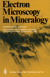
Electron Microscopy in Mineralogy
Herausgegeben von Wenk, H.-R.; Mitarbeit: Champness, P. E.; Christie, J. M.; Cowley, J. M.; Heuer, A. H.; Thomas, G.; Tighe, N. J.
Softcover reprint of the original 1st ed. 1976. 2011. xiv, 566 S. XII, 564 pp. 272 figs., 15 tabs. 244
Verlag/Jahr: SPRINGER, BERLIN 2011
ISBN: 3-642-66198-X (364266198X)
Neue ISBN: 978-3-642-66198-3 (9783642661983)
Preis und Lieferzeit: Bitte klicken
During the last five years transmission electron microscopy (TEM) has added numerous important new data to mineralogy and has considerably changed its outlook. This is partly due to the fact that metallurgists and crystal physicists having solved most of the structural and crystallographic problems in metals have begun to show a widening interest in the much more complicated structures of minerals, and partly to recent progress in experimental techniques, mainly the availability of ion-thinning devices. While electron microscopists have become increasingly interested in minerals (judging from special symposia at recent meetings such as Fifth European Congress on Electron microscopy, Man chester 1972; Eight International Congress on Electron Microscopy, Canberra 1974) mineralogists have realized advantages of the new technique and applied it with increasing frequency. In an effort to coordinate the growing quantity of research, electron microscopy sessions have been included in meetings of mineralogists (e. g. Geological Society of America, Minneapolis, 1972, American Crystallographic Association, Berkeley, 1974). The tremendous response for the TEM symposium which H. -R. Wenk and G. Thomas organized at the Berkeley Conference of the American Crystallographic Association formed the basis for this book. It appeared useful at this stage to summarize the achievements of electron microscopy, scattered in many different journals in several different fields and present them to mineralogists. A group of participants as the Berkeley symposium formed an Editorial Committee and outlined the content of this book.
Section 1 Introduction.- Section 2 Contrast.- 2.1 Fundamentals of Electron Microscopy.- 2.2 Interpretation of Electron Diffraction Patterns.- 2.3 Contrast Effects at Planar Interfaces.- 2.4 Computer Simulation of Dislocation Images in Quartz.- 2.5 The Direct Imaging of Crystal Structures.- 2.6 A Comparison of Bright Field and Dark Field Imaging of Pyrrhotite Structures.- Section 3 Experimental Techniques.- Section 4 Exsolution.- 4.1 Exsolution in Silicates.- 4.2 Coarsening in a Spinodally Decomposing System: TiO2-SnO2.- 4.3 Magnetite Lamellae in Reduced Hematites.- 4.4 Precipitation in the Ilmenite-hematite System.- 4.5 Pigeonite Exsolution from Augite.- 4.6 The Transformation of Pigeonite to Orthopyroxene.- 4.7 On the Detailed Structure of Ledges in an Augite-enstatite Interface.- 4.8 The Phase Distributions in Some Exsolved Amphiboles.- 4.9 Physical Aspects of Exsolution in Natural Alkali Feldspars.- 4.10 Analytical Electron Microscopy of Exsolution Lamellae in Plagioclase Feldspars.- 4.11 Exsolution in Metamorphic Bytownite.- Section 5 Polymorphic Phase Transitions.- 5.1 Polymorphic Phase Transitions in Minerals.- 5.2 Direct Observation of Iron Vacancies in Polytypes of Pyrrhotite.- 5.3 Rutile: Planar Defects and Derived Structures.- 5.4 High-resolution Electron Microscopy of Unit Cell Twinning in Enstatite.- 5.5 Polytypism in Wollastonite.- 5.6 High-resolution Electron Microscopy of Labradorite Feldspar.- 5.7 Origin of the (c) Domains of Anorthite.- 5.8 On Polymorphism of BaAl2Si2O8.- 5.9 The Submicroscopic Structure of Wenkite.- Section 6 Deformation Defects.- 6.1 Deformation Structures in Minerals.- 6.2 Work Hardening and Creep Deformation of Corundum Single Crystals.- 6.3 Dislocation Structures in Synthetic Quartz.- 6.4 The Microstructure of Some Naturally Deformed Quartzites.- 6.5 Defects in Deformed Calcite and Carbonate Rocks.- 6.6 Plasticity of Olivine in Peridotites.- 6.7 The Role of Crystal Defects in the Shear-induced Transformation of Orthoenstatite to Clinoenstatite.- Section 7 Special Techniques and Applications.- 7.1 Amorphous Materials.- 7.2 Signals Excited by the Scanning Beam.- 7.3 Analytical Electron Microscopy of Minerals.- 7.4 X-ray Microanalysis Using a Scanning Electron Microscope.- 7.5 Quantitative X-ray Microanalysis of Thin Foils.- 7.6 Particle Track Studies.- 7.7 Stony Meteorites.- 7.8 Microcracks in Crystalline Rocks.


