 Neuerscheinungen 2018Stand: 2020-02-01 |
Schnellsuche
ISBN/Stichwort/Autor
|
Herderstraße 10
10625 Berlin
Tel.: 030 315 714 16
Fax 030 315 714 14
info@buchspektrum.de |

Horst E. König, Hans-Georg Liebich
(Beteiligte)
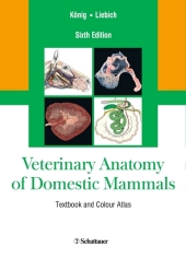
Veterinary Anatomy of Domestic Mammals
Textbook and Colour Atlas
Ed. by Horst E. König and Hans-Georg Liebich
6th rev. and extended ed. 2018. 824 S. 1089 Abb., 53 Tabellen. 300 mm
Verlag/Jahr: SCHATTAUER 2018
ISBN: 3-7945-2485-3 (3794524853) / 3-7945-2677-5 (3794526775) / 3-7945-2833-6 (3794528336)
Neue ISBN: 978-3-7945-2485-3 (9783794524853) / 978-3-7945-2677-2 (9783794526772) / 978-3-7945-2833-2 (9783794528332)
Preis und Lieferzeit: Bitte klicken
The standard atlas and textbook combination for students and practitioners alike!
Veterinary medicine is subject to constant change: The focus in veterinary schools is shifting more and more to real-life situations. In addition to basic anatomical knowledge and standard techniques, veterinary practitioners and surgeons need to keep abreast by obtaining specialized knowledge, including new imaging techniques.
This updated and expanded 6th edition of "Veterinary Anatomy of Domestic Mammals" will get you ready for both veterinary exams and clinical practice and will keep you at the cutting edge.
Excellent didactic graphs and clear structure make both studying and referencing fun! Numerous new, brilliant figures, especially on imaging techniques, bring anatomy to life and help you get a comprehensible grasp of clinical examination techniques.
- Unique text-atlas combination: anatomical basics and clinical specials in catchy combination with outstanding photographs and detailed graphics
- Approx. 1,100 figures: macroanatomical and histologic preparations, sliced plastinations, modern imaging techniques and additional coloured schemas
- New in the 6th edition:Additional chapter: "Sectional anatomy and imaging processes" with an introduction to plastination, CT, MRT, ultrasound and endoscopy, with a total of 51 new figures
- Added clinical examination techniques: rectal examination, equine endoscopy, palpation of bony structures
Introduction and general anatomy
Axial skeleton (skeleton axiale)
Fasciae and muscles of the head, neck and trunk
Forelimb or thoracic limb (membra thoracica)
Hindlimb or pelvic limb (membra pelvina)
Statics and dynamics
Body cavities and viscera
Digestive system (apparatus digestorius)
Respiratory system (apparatus respiratorius)
Urinary system (organa urinaria)
Male genital organs (organa genitalia masculina)
Female genital organs (organa genitalia feminina)
Organs of the cardiovascular system (systema cardiovasculare)
Immune system and lymphatic organs (organa lymphopoetica)
Nervous system (systema nervosum)
Endocrine glands (glandulae endocrinae)
Eye (organum visus)
Vestibulocochlear organ (organum vestibulocochleare)
Common integument (integumentum commune)
Topographical-clinical anatomy
Combining meticulous science and superb colour illustrations, this is a life-long source of reference for veterinary students, practitioners, educators and researchers, focusing on anatomical relationships to clinical conditions and where appropriate, to microscopic anatomy, histology, embryology and physiology.
From the contents:
General introduction
1 Axial skeleton
2 Fasciae and muscles of the head and trunk
3 Forelimb or thoracic limb
4 Hindlimb or pelvic limb
5 Statics and dynamics
6 Body cavities
7 Digestive system
8 Respiratory system
9 Urinary system
10 Male genital organs
11 Female genital organs
12 Organs of the cardiovascular system
13 Immune system and lymphatic organs
14 Nervous system
15 Endocrine glands
16 Eye
17 Vestibulocochlear organ
18 Common integument
19 Topographic clinical anatomy
References
Glossary of Terms
Index


