 Neuerscheinungen 2019Stand: 2020-02-01 |
Schnellsuche
ISBN/Stichwort/Autor
|
Herderstraße 10
10625 Berlin
Tel.: 030 315 714 16
Fax 030 315 714 14
info@buchspektrum.de |

Salvador Martinez, Margaret Martinez-de-la-Torre, Luis Puelles
(Beteiligte)
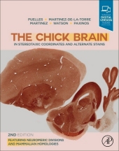
The Chick Brain in Stereotaxic Coordinates and Alternate Stains
Featuring Neuromeric Divisions and Mammalian Homologies
2. Aufl. 2019. 322 S. 276 mm
Verlag/Jahr: ACADEMIC PRESS 2019
ISBN: 0-12-566651-9 (0125666519) / 0-12-816040-3 (0128160403)
Neue ISBN: 978-0-12-566651-0 (9780125666510) / 978-0-12-816040-4 (9780128160404)
Preis und Lieferzeit: Bitte klicken
This atlas - and its accompanying text - is the most comprehensive work on avian neuroanatomy available so far. It identifies more than 900 hundred structures (versus ca. 250 in previous avian atlases), 180 of them for the first time. It correlates avian and mammalian neuroanatomy on the basis of homologies and applies mammalian terms to homologous avian structures. This is the first atlas that represents the fundamental histogenetic domains of the vertebrate neuroaxis on the basis of sound fate-mapping and gene expression data. This results in a substantial increase in accuracy of delineations. Developmental molecular biologists will find it easier to extrapolate early neural tube patterns into mature structures. The modern trend to shift avian neuroanatomical nomenclature toward mammalian terminology by reference to postulated homologies has been expanded to the entire brain, but is not yet complete. This creates a new standard for comparative cross-reference, which can also be applied to reptilian-mammalian comparisons.
Color photographs and matching diagrams of 65 coronal, 23 sagittal and 9 horizontal 140 micron-thick sections reacted histochemically for acetylcholinesterase (AChE).
Thoroughly revised drawings. Updated view of the pallium, including the new concept of homology between the lateral pallium and the mammalian claustro-insular complex.
Extensive introductory text and bibliography, presenting the background information, methodology and justification of delineations.
For the first time in any species, this atlas depicts the fate-mapped natural embryonic boundaries in the postnatal brain. For the first time, we present color images of all the 6 histological stains (AChE, Nissl, TH, calbindin, calretinin and parvalbumin) on which delineations are based (accompanying Expert Consult eBook).
Includes the Expert Consult eBook version, compatible with PC, Mac, and most mobile devices and eReaders, which allows readers to browse, search, and interact with content.
The eBook also contains annotatable AI files of diagrams for use by researchers.
1. Generalities, procedures and background information
2. Rationale for names applied in the atlas
3. Literature cited
4. List of abbreviations and explanations
5. Brain structures classified topographically by regions
Watson, Charles
Charles Watson is a neuroscientist and public health physician. His qualifications included a medical degree (MBBS) and two research doctorates (MD and DSc). He is Professor Emeritus at Curtin University, and holds adjunct professorial research positions at the University of New South Wales, the University of Queensland, and the University of Western Australia.
He has published over 100 refereed journal articles and 40 book chapters, and has co-authored over 25 books on brain and spinal cord anatomy. The Paxinos Watson rat brain atlas has been cited over 80,000 times. His current research is focused on the comparative anatomy of the hippocampus and the claustrum.
He was awarded the degree of Doctor of Science by the University of Sydney in 2012 and received the Distinguished Achievement Award of the Australasian Society for Neuroscience in 2018.


