 Neuerscheinungen 2017Stand: 2020-02-01 |
Schnellsuche
ISBN/Stichwort/Autor
|
Herderstraße 10
10625 Berlin
Tel.: 030 315 714 16
Fax 030 315 714 14
info@buchspektrum.de |

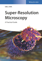
Udo J. Birk
Super-Resolution Microscopy
A Practical Guide
1. Auflage. 2017. 408 S. 133 SW-Abb., 24 Farbabb., 9 Tabellen. 244 mm
Verlag/Jahr: WILEY-VCH 2017
ISBN: 3-527-34133-1 (3527341331)
Neue ISBN: 978-3-527-34133-7 (9783527341337)
Preis und Lieferzeit: Bitte klicken
This unique book on super-resolution microscopy techniques presents comparative, in-depth analyses of the strengths and weaknesses of the individual approaches. It was written for non-experts who need to understand the principles of super-resolution or who wish to use recently commercialized instruments as well as for professionals who plan to realize novel microscopic devices. Explaining the practical requirements in terms of hardware, software and sample preparation, the book offers a wealth of hands-on tips and practical tricks to get a setup running, provides invaluable help and support for successful data acquisition and specific advice in the context of data analysis and visualization. Furthermore, it addresses a wide array of transdisciplinary fields of applications.
The author begins by outlining the joint efforts that have led to achieving super-resolution microscopy combining advances in single-molecule photo-physics, fluorophore design and fluorescent labeling, instrument design and software development. The following chapters depict and compare current main standard techniques such as structured illumination microscopy, single-molecule localization, stimulated emission depletion microscopy and multi-scale imaging including light-sheet and expansion microscopy. For each individual approach the experimental setups are introduced, the imaging protocols are provided and the various applications illustrated. The book concludes with a discussion of future challenges addressing issues of routine applications and further commercialization of the available methods.
Guiding users in how to make choices for the design of their own experiments from scratch to promising application, this one-stop resource is intended for researchers in the applied sciences, from chemistry to biology and medicine to physics and engineering.
Preface
Abbreviations
INTRODUCTION
The Classical Resolution Limit
Methods to Circumvent the Classical Resolution Barrier in Fluorescence Microscopy
Implementation of Super-Resolution Microscopy (SRM)
Contrast
Applications to Study Nuclear DNA
Other Applications
PHYSICOCHEMICAL BACKGROUND
Motivation
Labeling
Transitions of the Fluorophores
Samples
HARD- AND SOFTWARE
Hardware Requirements
Software
Open Source and Best Practice
STRUCTURED ILLUMINATION AND IMAGE SCANNING MICROSCOPY
Axially Structured Illumination Microscopy (aSIM)
Laterally Structured Illumination Microscopy (SIM)
Image Scanning Microscopy
Super-Resolution Using Rotating Coherent Scattering (ROCS) Microscopy
LOCALIZATION MICROSCOPY
On the Principles of Localization Microscopy
PALM/STORM/fPALM/SPDM Approach
Implementation of SMLM
On the Principles of Three-Dimensional SMLM
Reduction of Out-of-Focus Light
How to Build a Three-Dimensional SMLM
High-Density Single Emitter Microscopy Methods: SOFI, 3B, SHRImP, etc.
Approaches Towards Counting Molecules
Requirements and Sample Preparation
Data Acquisition
Data Analysis
Troubleshooting
Meta Analysis Tailored for SMLM
Example Applications
STIMULATED EMISSION DEPLETION MICROSCOPY (STED)
On the Principles of Stimulated Emission Depletion Microscopy
Implementation of STED
Fluorescent Probes
Dye Combinations for Dual-Color STED
Requirements and Sample Preparation
Data Acquisition
Data Analysis and Visualization
Example Applications
Conclusion
MULTI-SCALE IMAGING
Light-Sheet Fluorescence Microscopy (LSFM)
Optical Projection Tomography (OPT)
Expansion Microscopy (ExM) and Sample Clearing
Alternative Approaches
DISCUSSION
Future Challenges
Commercialization of Super-Resolution Microscopes
Concluding Remarks
Index
Udo Birk studied physics and mathematics in Canada and Germany and obtained a PhD in applied physics from the University of Heidelberg (Germany) for his work on tissue spectrometry and spatially modulated illumination microscopy. He was a Marie-Curie Fellow at King´s College London (UK) and at the Foundation of Research and Technology Hellas (Greece), where he worked on structured illumination microscopy and on optical projection tomography. Currently, he is deputy group leader at the Institute of Molecular Biology in Mainz (Germany), which equals a position of an associate professor, and his research focuses on the development and application of advanced optical imaging techniques.


